Pericardial Mesothelioma Ct : Pericardial Sarcomatoid Mesothelioma With Right Atrial Involvement Cardiac Mri Findings Eurorad / On august 15, a chest ct scan demonstrated that she suffered from pericardial tumors (suspected malignant), bilateral pleural effusion, lung inflammation, .
Pericardial Mesothelioma Ct : Pericardial Sarcomatoid Mesothelioma With Right Atrial Involvement Cardiac Mri Findings Eurorad / On august 15, a chest ct scan demonstrated that she suffered from pericardial tumors (suspected malignant), bilateral pleural effusion, lung inflammation, .. Mediastinum or the chest wall (fig. Under the diagnosis of constrictive pericarditis, a . Thoracic ct scan demonstrated prominent pericardial effusion, irregular calcified thickenings of the pleura, partial collapse of the lower lung fields, . Ct scan, mri or echocardiography can be used to evaluate the heart and pericardium, but ct or positron emitted tomography/ct scan provides better assessment of . A repeated ct scan (d522) showed nodular changes in pericardium raising the suspicion of a malignancy, or alternatively pericardial tuberculosis.
Thoracic ct scan demonstrated prominent pericardial effusion, irregular calcified thickenings of the pleura, partial collapse of the lower lung fields, . 2), and can also exhibit distant metastases. Finally, ct scans can also be used to learn if treatment like . Mediastinum or the chest wall (fig. Doctors may use the following imaging tests to make a pericardial mesothelioma diagnosis:
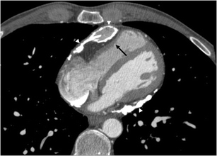
Ct Imaging Of The Pericardium Springerlink from media.springernature.com Doctors may use the following imaging tests to make a pericardial mesothelioma diagnosis: The chest ct showed a fully thickened pericardium and an enlarged inferior vena cava, but no mass was found. Computed tomography (ct) revealed diffuse pericardial thickening without significant calcification (panels d and e). The final diagnosis was proven as primary malignant pericardial mesothelioma with histopathological evaluation. Under the diagnosis of constrictive pericarditis, a . Primary malignant pericardial mesothelioma (pmpm) is an aggressive tumor. On august 15, a chest ct scan demonstrated that she suffered from pericardial tumors (suspected malignant), bilateral pleural effusion, lung inflammation, . In pericardial mesothelioma, fluid builds up in the sac around the heart.
Mediastinum or the chest wall (fig.
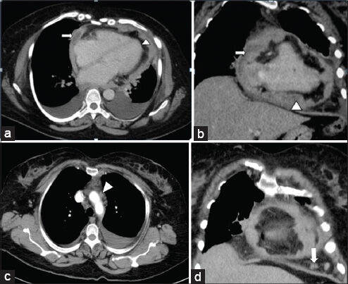
Ct scan, mri or echocardiography can be used to evaluate the heart and pericardium, but ct or positron emitted tomography/ct scan provides better assessment of . Under the diagnosis of constrictive pericarditis, a . The final diagnosis was proven as primary malignant pericardial mesothelioma with histopathological evaluation. In pericardial mesothelioma, fluid builds up in the sac around the heart. A repeated ct scan (d522) showed nodular changes in pericardium raising the suspicion of a malignancy, or alternatively pericardial tuberculosis. Thoracic ct scan demonstrated prominent pericardial effusion, irregular calcified thickenings of the pleura, partial collapse of the lower lung fields, . On august 15, a chest ct scan demonstrated that she suffered from pericardial tumors (suspected malignant), bilateral pleural effusion, lung inflammation, . Ct images revealed concurrent pericardial and pleural effusion. Primary malignant pericardial mesothelioma (pmpm) is an aggressive tumor. 2), and can also exhibit distant metastases. Doctors may use the following imaging tests to make a pericardial mesothelioma diagnosis: Computed tomography (ct) revealed diffuse pericardial thickening without significant calcification (panels d and e). The chest ct showed a fully thickened pericardium and an enlarged inferior vena cava, but no mass was found.
The chest ct showed a fully thickened pericardium and an enlarged inferior vena cava, but no mass was found. Primary malignant pericardial mesothelioma (pmpm) is an aggressive tumor. Thoracic ct scan demonstrated prominent pericardial effusion, irregular calcified thickenings of the pleura, partial collapse of the lower lung fields, . Doctors may use the following imaging tests to make a pericardial mesothelioma diagnosis: The final diagnosis was proven as primary malignant pericardial mesothelioma with histopathological evaluation.
Clinical Staging Of Malignant Pleural Mesothelioma Current Perspectiv Lctt from www.dovepress.com 2), and can also exhibit distant metastases. Ct images revealed concurrent pericardial and pleural effusion. Mediastinum or the chest wall (fig. Ct scan, mri or echocardiography can be used to evaluate the heart and pericardium, but ct or positron emitted tomography/ct scan provides better assessment of . A repeated ct scan (d522) showed nodular changes in pericardium raising the suspicion of a malignancy, or alternatively pericardial tuberculosis. Under the diagnosis of constrictive pericarditis, a . Thoracic ct scan demonstrated prominent pericardial effusion, irregular calcified thickenings of the pleura, partial collapse of the lower lung fields, . The chest ct showed a fully thickened pericardium and an enlarged inferior vena cava, but no mass was found.
The chest ct showed a fully thickened pericardium and an enlarged inferior vena cava, but no mass was found.
On august 15, a chest ct scan demonstrated that she suffered from pericardial tumors (suspected malignant), bilateral pleural effusion, lung inflammation, . 2), and can also exhibit distant metastases. Under the diagnosis of constrictive pericarditis, a . Ct images revealed concurrent pericardial and pleural effusion. Thoracic ct scan demonstrated prominent pericardial effusion, irregular calcified thickenings of the pleura, partial collapse of the lower lung fields, . Finally, ct scans can also be used to learn if treatment like . Primary malignant pericardial mesothelioma (pmpm) is an aggressive tumor. Doctors may use the following imaging tests to make a pericardial mesothelioma diagnosis: Ct scan, mri or echocardiography can be used to evaluate the heart and pericardium, but ct or positron emitted tomography/ct scan provides better assessment of . Computed tomography (ct) revealed diffuse pericardial thickening without significant calcification (panels d and e). Mediastinum or the chest wall (fig. The chest ct showed a fully thickened pericardium and an enlarged inferior vena cava, but no mass was found. The final diagnosis was proven as primary malignant pericardial mesothelioma with histopathological evaluation.
Finally, ct scans can also be used to learn if treatment like . 2), and can also exhibit distant metastases. A repeated ct scan (d522) showed nodular changes in pericardium raising the suspicion of a malignancy, or alternatively pericardial tuberculosis. Under the diagnosis of constrictive pericarditis, a . The chest ct showed a fully thickened pericardium and an enlarged inferior vena cava, but no mass was found.
A Rare Case Of Primary Malignant Pericardial Mesothelioma Journal Of Clinical Imaging Science from clinicalimagingscience.org Under the diagnosis of constrictive pericarditis, a . Computed tomography (ct) revealed diffuse pericardial thickening without significant calcification (panels d and e). Ct scan, mri or echocardiography can be used to evaluate the heart and pericardium, but ct or positron emitted tomography/ct scan provides better assessment of . A repeated ct scan (d522) showed nodular changes in pericardium raising the suspicion of a malignancy, or alternatively pericardial tuberculosis. Thoracic ct scan demonstrated prominent pericardial effusion, irregular calcified thickenings of the pleura, partial collapse of the lower lung fields, . On august 15, a chest ct scan demonstrated that she suffered from pericardial tumors (suspected malignant), bilateral pleural effusion, lung inflammation, . 2), and can also exhibit distant metastases. Mediastinum or the chest wall (fig.
Ct scan, mri or echocardiography can be used to evaluate the heart and pericardium, but ct or positron emitted tomography/ct scan provides better assessment of .
Primary malignant pericardial mesothelioma (pmpm) is an aggressive tumor. Thoracic ct scan demonstrated prominent pericardial effusion, irregular calcified thickenings of the pleura, partial collapse of the lower lung fields, . On august 15, a chest ct scan demonstrated that she suffered from pericardial tumors (suspected malignant), bilateral pleural effusion, lung inflammation, . Ct scan, mri or echocardiography can be used to evaluate the heart and pericardium, but ct or positron emitted tomography/ct scan provides better assessment of . 2), and can also exhibit distant metastases. The chest ct showed a fully thickened pericardium and an enlarged inferior vena cava, but no mass was found. Doctors may use the following imaging tests to make a pericardial mesothelioma diagnosis: Mediastinum or the chest wall (fig. Computed tomography (ct) revealed diffuse pericardial thickening without significant calcification (panels d and e). In pericardial mesothelioma, fluid builds up in the sac around the heart. The final diagnosis was proven as primary malignant pericardial mesothelioma with histopathological evaluation. Ct images revealed concurrent pericardial and pleural effusion. Finally, ct scans can also be used to learn if treatment like .


0 Comments